Warning: include_once(/home/u171827962/domains/drnehatiwarieyeclinic.com/public_html/wp-content/plugins/elementor-pro/libraries/plugins.php): Failed to open stream: No such file or directory in /home/u171827962/domains/drnehatiwarieyeclinic.com/public_html/wp-content/plugins/elementor-pro/elementor-pro.php on line 15

Warning: include_once(): Failed opening '/home/u171827962/domains/drnehatiwarieyeclinic.com/public_html/wp-content/plugins/elementor-pro/libraries/plugins.php' for inclusion (include_path='.:/opt/alt/php81/usr/share/pear:/opt/alt/php81/usr/share/php:/usr/share/pear:/usr/share/php') in /home/u171827962/domains/drnehatiwarieyeclinic.com/public_html/wp-content/plugins/elementor-pro/elementor-pro.php on line 15
Retina & Fundus Examination - Dr. Neha Tiwari
Ophthalmologist Preloader

Dr. Neha Tiwari is a dedicated and compassionate General Ophthalmologist with extensive experience in providing comprehensive eye care services.

Visiting Hours

Gallery Posts

Retina & Fundus Examination

Retina and Fundus Examination

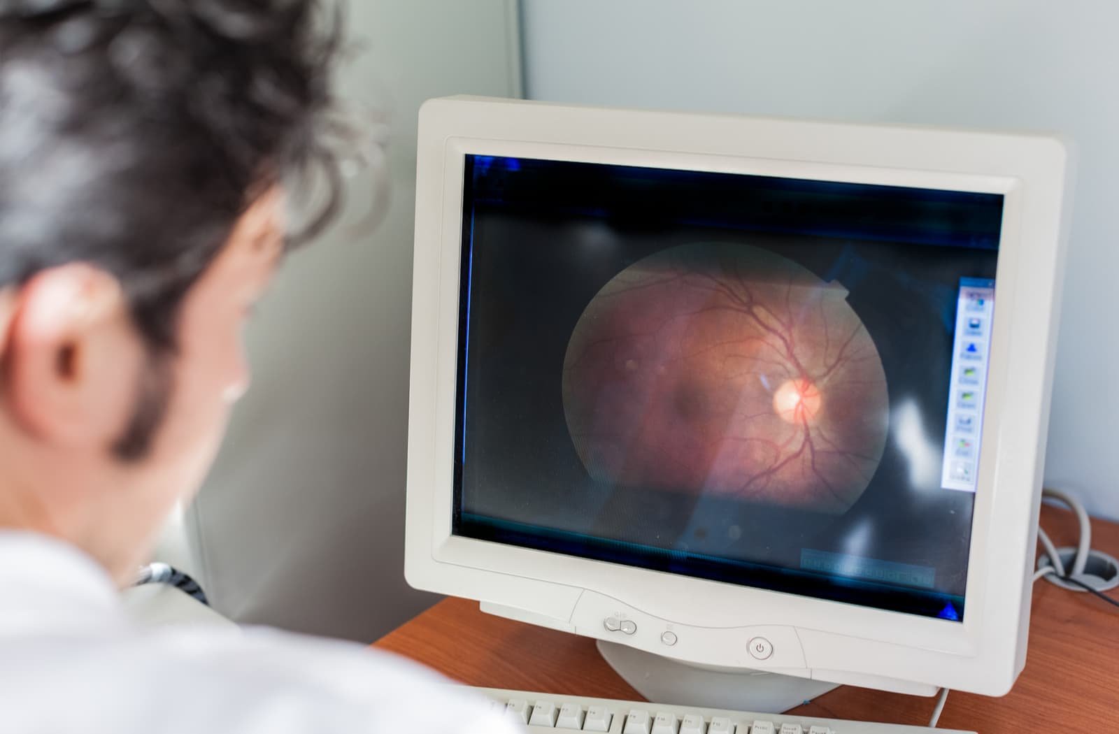
Retina and Fundus Examination are critical diagnostic procedures used to evaluate the health of the eye’s interior structures, particularly the retina, optic disc, macula, and blood vessels. These exams help in detecting and managing various eye conditions that can affect vision.
What is a Retina Examination?

  • A retina examination involves evaluating the light-sensitive layer at the back of the eye, known as the retina. The retina is crucial for vision as it converts light into electrical signals that are sent to the brain.
  • It is used to detect conditions such as diabetic retinopathy, age-related macular degeneration (AMD), retinal detachment, and other retinal disorders.

2. What is a Fundus Examination?

  • The term “fundus” refers to the interior surface of the eye, including the retina, optic disc, macula, and retinal blood vessels.
  • A fundus examination, also called ophthalmoscopy, allows the doctor to view and assess these structures to detect signs of disease or damage.

3. Types of Retina and Fundus Examinations

  • Direct Ophthalmoscopy : This uses a handheld device with a light to directly visualize the back of the eye. It gives a limited view but is quick and useful for basic assessment.
  • Indirect Ophthalmoscopy : In this method, the doctor uses a head-mounted light and a handheld lens to examine a larger area of the retina. It provides a more comprehensive view and is often used for diagnosing retinal detachment.
  • Fundus Photography : This involves taking a photograph of the interior surface of the eye. It helps in documenting the condition of the retina and tracking changes over time.
  • Optical Coherence Tomography (OCT) : An advanced imaging technique that provides detailed cross-sectional images of the retina. It is especially useful for detecting conditions like macular edema and AMD.
  • Fluorescein Angiography : A diagnostic procedure where a fluorescent dye is injected into a vein, and images are taken to evaluate the blood flow in the retina’s blood vessels. It helps diagnose diabetic retinopathy and other vascular conditions.

4. Conditions Detected by Retina and Fundus Examinations

  • Diabetic Retinopathy : Changes in the retina’s blood vessels caused by diabetes.
  • Age-Related Macular Degeneration (AMD) : Affects the central part of the retina, leading to vision loss.
  • Glaucoma : Characterized by damage to the optic nerve, often due to increased eye pressure.
  • Retinal Detachment : The retina separates from the back of the eye, which can cause permanent vision loss if not treated promptly.
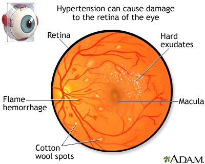
  • Hypertensive Retinopathy : Changes in the retina due to high blood pressure.

5. Procedure for Retina and Fundus Examination

  • Preparation : The eye doctor may dilate the pupils using special eye drops to get a better view of the retina. This may cause blurry vision and light sensitivity for a few hours.
  • Examination : The doctor uses specialized tools like an ophthalmoscope or slit-lamp microscope with a lens to view the eye’s interior.
  • Post-Examination Care : Patients may need to wear sunglasses to protect their eyes from bright light until the dilation wears off.

6. Importance of Regular Retina and Fundus Examinations

  • These exams are essential for detecting eye diseases early, especially conditions like diabetic retinopathy or AMD, which may not show symptoms in the early stages.
  • Early detection through regular examinations can help manage conditions effectively and prevent significant vision loss.

Retina and fundus examinations play a vital role in maintaining eye health and preserving vision. Regular eye check-ups, especially for people with risk factors like diabetes or hypertension, are important for early detection and treatment of potential eye conditions.

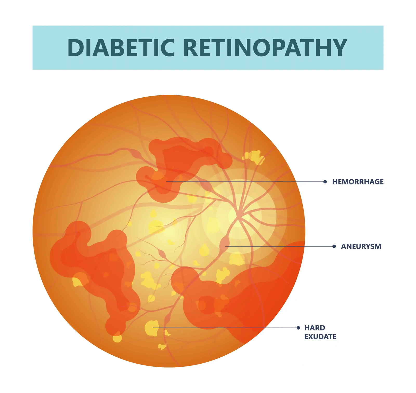
Diabetic Retinopathy

Diabetic Retinopathy is a complication of diabetes that affects the eyes. It occurs when high blood sugar levels damage the blood vessels in the retina, the light-sensitive tissue at the back of the eye. This condition can lead to vision problems and, if left untreated, may result in blindness.
Symptoms :

  • Blurred vision
  • Floaters or dark spots
  • Difficulty seeing colors
  • Sudden vision loss (in severe cases)

Prevention and Management :

  • Control blood sugar, blood pressure, and cholesterol : Helps reduce the risk.
  • Regular eye exams : Early detection can prevent serious vision problems.
  • Treatment options:
    • Laser therapy : To stop or slow blood leakage.
    • Anti-VEGF injections : To reduce swelling and inhibit abnormal blood vessel growth.
    • Vitrectomy surgery : In severe cases, to remove blood or scar tissue.

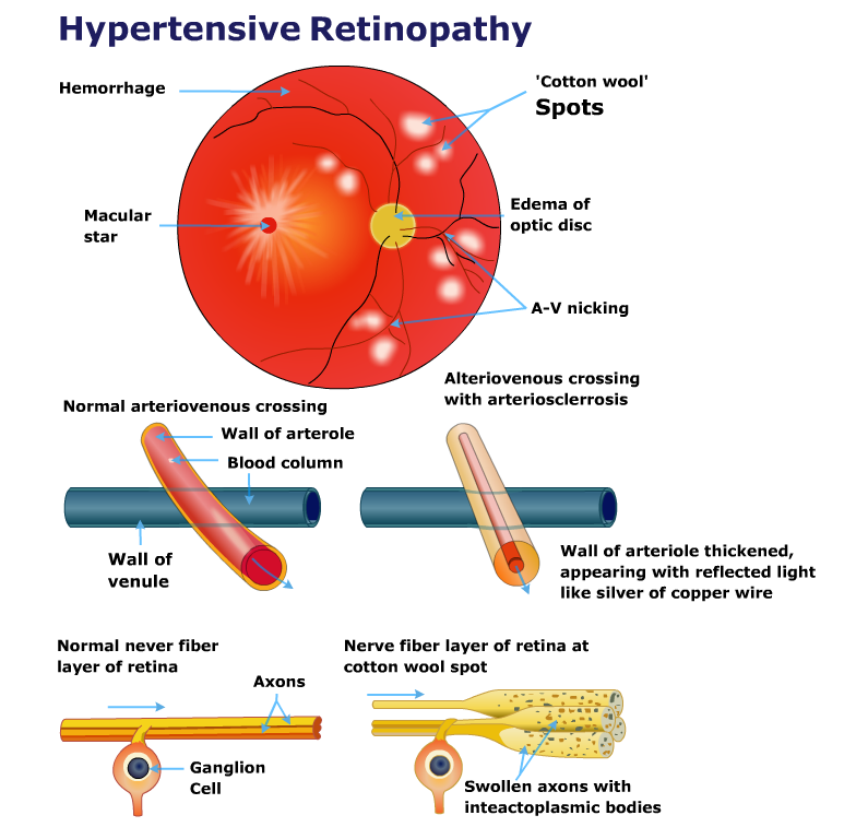
Hypertensive Retinopathy

Hypertensive Retinopathy is an eye condition that occurs due to long-term high blood pressure (hypertension). Elevated blood pressure damages the blood vessels in the retina, the light-sensitive layer at the back of the eye, potentially leading to vision problems.

Symptoms :

  • Often asymptomatic in early stages
  • Blurred vision
  • Double vision
  • Headaches
  • Sudden vision loss (in advanced cases)

Prevention and Management :

  • Control blood pressure : Maintaining normal blood pressure levels can help prevent or slow the progression.
  • Healthy lifestyle : Eating a balanced diet, exercising, reducing salt intake, and avoiding smoking.
  • Regular eye exams : Early detection can help manage the condition before severe damage occurs.

Hypertensive retinopathy F&Q's

Hypertensive retinopathy is a condition where high blood pressure causes damage to the blood vessels in the retina, potentially leading to vision problems.

The main cause is long-term, uncontrolled high blood pressure, which puts excessive pressure on the small blood vessels in the eyes, causing damage and changes in the retina.

Symptoms may include blurred vision, headaches, double vision, reduced vision, and in severe cases, sudden loss of vision. However, some individuals may not experience any symptoms until the condition has progressed significantly.

An eye doctor (ophthalmologist) can diagnose hypertensive retinopathy during a comprehensive eye exam. The examination may include retinal imaging or fundoscopy to check for signs of damage to the retinal blood vessels.

Yes, if left untreated, severe hypertensive retinopathy can lead to significant vision loss or even blindness due to damage to the retina.

The primary treatment is managing blood pressure through lifestyle changes, medication, and regular monitoring. In advanced cases, laser treatment or other procedures may be required to manage retinal damage.